
Virtual Rounds Session 2: Pneumothorax (Premed Shadowing)
4 min read
Skip To:
Throughout each premed virtual shadowing session, you will actively participate by answering and asking questions about the week’s proposed case. Like an in-person shadowing experience, we strive to stimulate students intellectually and ultimately reaffirm their interest in medicine.
Introduction – Tension Pneumothorax
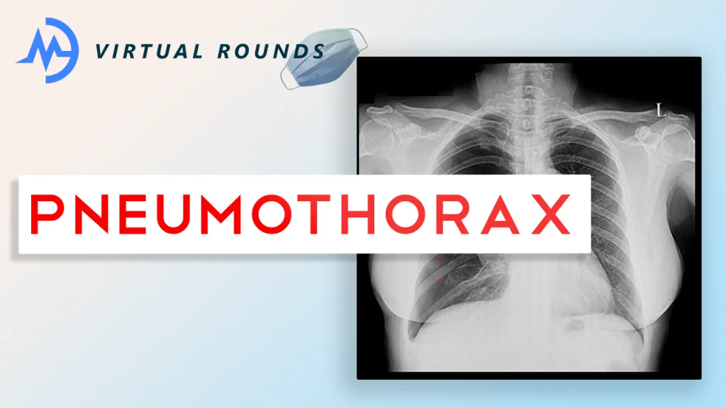
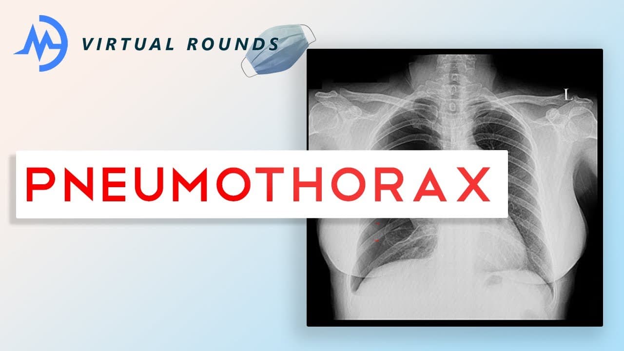
If you’re interested in emergency medicine, then this will be a fascinating topic for you! Tension pneumothorax is a common pathology encountered in traumatic situations, such as a car accident or stab wound to the chest. It is a life-threatening emergency that requires an immediate clinical diagnosis in order to save the patient’s life. Follow along with the case below to learn how to recognize, diagnose, and save a patient with a tension pneumothorax!
Case Highlights
- A 22-year-old male presents to the emergency department by ambulance with a chief complaint of a stab wound to the right chest. As the patient is wheeled into the trauma bay, you observe that he is unconscious and the knife is still in place. The EMS (emergency medical service) personnel inform you that the patient recently lost consciousness. As the medical team gets him hooked up to the monitor and you start your primary survey (airway, breathing, circulation), you realize that he is becoming hypoxemic (low blood oxygen saturation leading to suffocation) and suffering from cardiorespiratory compromise (low blood pressure, the heart not able to pump effectively).
- As an emergency resident physician, you use your clinical judgement to determine that the patient needs immediate treatment in order to survive. You notice that he has distended neck veins (sign of high intrathoracic pressure), tachycardia (pulse over 100 beats per minute), absent breath sounds and hyperresonance on the right side where he was stabbed. Before performing the lifesaving procedure, you take only a few seconds to share your differential diagnosis with your attending to ensure you are making the right decision…
Pathology
Under normal circumstances, where a patient does not require immediate diagnosis and treatment, it is important to stop and consider your differential diagnosis (the other diseases/conditions that a patient could also have given his/her symptoms and physical exam). We want to be able to determine what the patient has before treating them, so it is important to acknowledge the other possibilities and then rule them out. For our patient, we want to consider the following:
Tension Pneumothorax
This condition occurs due to the creation of a one-way valve that lets air into the chest but does not let it out. With each breath, the pressure within the chest increases and puts tremendous pressure on the lung(s), heart and great vessels (inferior and superior vena cava). The reason we believe that the patient has a tension pneumothorax is due to our physical exam findings and vital signs. Due to the patient’s lack of breath sounds and hyperresonance on the right side, the patient likely has a collapsed lung (that is what pneumothorax means). Due to the very patient’s low blood pressure, distended neck veins, and hypoxia, it is likely that the patient has increasing pressure in his chest causing compression of his heart and left lung. The increasing pressure prevents blood from flowing into the heart (hence the distended neck veins) and leaving the heart (not able to fill all the way due to being squished by the increasing pressure in the chest). All of these findings convince us that the patient has a tension pneumothorax and must therefore be treated ASAP with needle decompression (creating a small hole so the air can escape the chest)
Pulmonary Embolism
A pulmonary embolism occurs due to a clot that can, for example, originate in a patient’s leg and travel up to his/her lung. Patient’s with a pulmonary embolism (PE) can present in a similar way to tension pneumothorax. They could have tachycardia, chest pain, and hypoxemia. However, we would not expect a patient to have absent breath sounds in either lung. Additionally, we would expect the patient to have pleuritic chest pain (pain with inhalation and exhalation) and a cough and/or hemoptysis (coughing up blood).
Cardiac Tamponade
It is important to consider this diagnosis as it can also occur due to traumatic situations like a car accident or car accident. Cardiac tamponade occurs when blood enters the pericardial space (the heart is covered by a sac called the pericardium) and causes compression of the heart itself, preventing it from circulating blood throughout the body. This leads to specific and similar exam findings, including low blood pressure, tachycardia, chest pain, and distended neck veins. A key physical exam finding to consider is muffled heart sounds (when you listen to the patient’s heart, it is more difficult to hear the individual beats). We would not expect a patient with cardiac tamponade to have absent breath sounds.
Take home points
- Tension pneumothorax occurs due to the creation of a one-way valve that leads to increasing pressure within the chest. It is a medical emergency that requires immediate treatment to save the patient’s life!
- Tension pneumothorax is a clinical diagnosis, meaning you can diagnose it based on vital signs (heart rate, blood pressure, oxygen saturation) and physical exam findings (distended neck veins, absent breath sounds/hyperresonance on the side of the collapsed lung, weak pulse).
- To save the patient’s life, you must perform a needle decompression in the second or third intercostal space (space between the ribs), which creates a small hole to let the collected air escape.
- Finally, make sure you consider emergency medicine as a potential specialty! You’ll have the most exciting career and be trained to handle any situation-you never know what’s coming through the front doors!
Watch the virtual shadowing session here:
Fill out this form throughout the session:
Written by Motivate MD